코미팜

목록목록
코미녹스 기전을 연구 뇌종양 mRNA 전사 억제효과는
  • 20/06/24 12:21
  • 조회 465
gregory16 자기 소개가 없습니다.


AJGP > 2020 > April > Current management of cerebral gliomas

Clinical
Volume 49, Issue 4, April 2020

Current management of cerebral gliomas

Rosalind L Jeffree   
doi: 10.31128/AJGP-09-19-5063   |    Download article
Cite this article    BIBTEX    REFER    RIS


AJGP> 2020> 4 월> 뇌 신경 교종의 현재 관리


객관적인
2049 년 4 호 49 호


뇌 신경 교종의 현재 관리
로잘린드 L 제프리
-------------------------------------------------- ------------------------------


doi : 10.31128 / AJGP-09-19-5063 | 기사 다운로드
이 기사를 인용하십시오 BIBTEX REFER RIS


Background

Despite their rarity, primary tumours of the central nervous system have a devastating impact on patient survival and quality of life. The classification of glial tumours has recently changed, and large trials have provided data on treatment impact; however, the treatment armamentarium remains the same, and many questions persist.

Objective

The aim of this narrative review is to discuss the current understanding and management of the most common glial brain tumours to equip general practitioners (GPs) and other non-neuro-oncological specialists with appropriate knowledge to share care and support patients.

Discussion

Treatment of brain tumours is complex and multifaceted, and it involves many different specialists. Recent advances in translational research and molecular understanding of brain tumours raise hope that new treatments are imminent, and patients should be encouraged to participate in clinical trials. The GP has an important role in patient support and coordination of care.


Although glioblastoma multiforme (GBM) is the most common primary brain tumour, it has an incidence of only 6.8 per 100,000. As a result, most general practitioners (GPs) will care for very few patients with this condition, and low-grade tumours are even less common.1 However, the devastating functional and social ramifications mean GBM confers a disproportionate burden to patients and GPs. The family doctor has an essential role in surveillance, support and coordination of care. Despite improvements in surgery, chemotherapy and radiotherapy, the prognosis remains poor for most patients with high-grade gliomas.

Presentation and initial investigation

Brain tumours present a significant diagnostic challenge for primary care physicians because early symptoms are non-specific. A recent large observational study identified headache as the most sensitive symptom but found that only two in 1000 patients with headache had a brain tumour. Similarly, only 1.6% of patients with adult-onset seizures have a tumour.2 Combinations of symptoms are more sensitive ? particularly headache, cognitive problems and weakness ? but still have a positive predictive value of under 10% (ie only one in 10 patients with headache and cognitive problems or headache and weakness will have a tumour). Observing temporal trends in symptoms and identifying cognitive issues through collateral history or simple testing may be the best way to identify patients with a tumour.2 By the time the patient develops classic symptoms of raised intracranial pressure and morning headache and vomiting, with or without neurological deficit, the situation is an emergency.

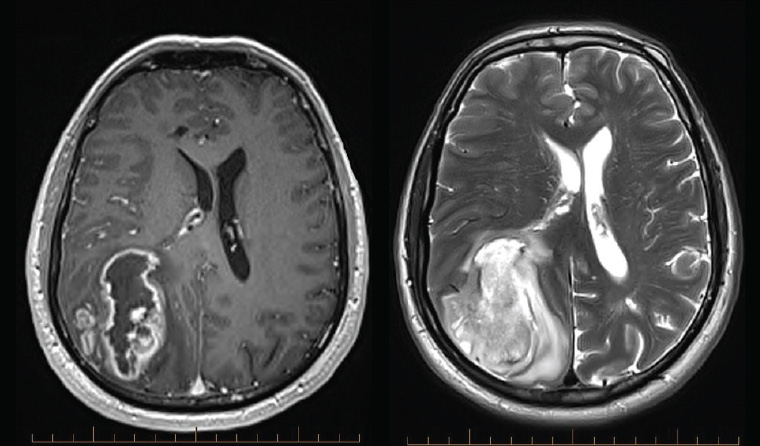
A magnetic resonance imaging (MRI) scan is the appropriate initial investigation to diagnose or exclude an intracranial lesion. The imaging appearance of GBM is often very characteristic, showing peripheral enhancement around a region of central necrosis with variable degrees of surrounding oedema (Figure 1). This is concerning because the differential diagnosis is metastatic cancer or abscess. Low-grade gliomas appear hypointense on T1 and hyperintense on T2 without the necrotic centre or enhancement (Figure 2). Newer imaging modalities such as positron-emission tomography (PET), MRI spectroscopy and perfusion imaging show promise but do not yet have the sensitivity or specificity to change usual management.3,4 Despite this, high uptake areas on fluorodeoxyglucose (FDG)-PET (18F-fluorodeoxy-glucose) or amino acid-based PET (eg 11C-methyl-methionine [11C-MET], 18F-fluorophenylalanine [18F-DOPA] or 18F-fluoroethyl-l-tyrosine [18F-FET])

배경

희귀 함에도 불구하고 중추 신경계의 주요 종양은 환자의 생존과 삶의 질에 치명적인 영향을 미칩니다. 신경아 교종의 분류는 최근에 변경되었으며, 대규모 시험에서 치료 영향에 대한 데이터가 제공되었습니다. 그러나, 치료 용 armamentarium은 동일하게 유지되며 많은 질문이 지속됩니다.


객관적인

이 내러티브 검토의 목적은 일반 개업의 (GP) 및 기타 비-신경 종양학 전문가에게 적절한 지식을 제공하여 치료를 공유하고 환자를 지원하는 가장 일반적인 신경교 뇌종양의 현재 이해 및 관리에 대해 논의하는 것입니다.


토론

뇌종양의 치료는 복잡하고 다면적이며 많은 다른 전문가들이 필요합니다. 최근 뇌종양의 번역 연구 및 분자 이해의 진보는 새로운 치료가 임박하고 환자가 임상 시험에 참여하도록 장려해야한다는 희망을 불러 일으킨다. GP는 환자 지원 및 치료 조정에 중요한 역할을합니다.


다형성 교 모세포종 (GBM)이 가장 흔한 1 차 뇌종양이지만, 100,000 명당 발생률은 6.8입니다. 결과적으로, 대부분의 일반의 (GP)는이 질환을 가진 환자를 거의 돌보지 않을 것이며, 낮은 등급의 종양은 훨씬 덜 일반적입니다 .1 그러나, 기능과 사회적 영향이 치명적이라는 것은 GBM이 환자와 GP에게 불균형적인 부담을 준다는 것을 의미합니다. 가정의는 진료의 감시, 지원 및 조정에 필수적인 역할을합니다. 수술, 화학 요법 및 방사선 요법의 개선에도 불구하고, 예후는 고급 신경 교종 환자의 대부분에게 좋지 않습니다.

발표 및 초기 조사

뇌종양은 초기 증상이 비특이적이므로 일차 진료 의사에게 심각한 진단 문제를 제시합니다. 최근의 대규모 관찰 연구에 따르면 두통이 가장 민감한 증상으로 밝혀졌지만 두통이있는 1000 명 중 2 명만이 뇌종양이있는 것으로 나타났습니다. 마찬가지로 성인 발작 환자의 1.6 %만이 종양을 가지고 있습니다 .2 증상의 조합은 더욱 민감합니다. 특히 두통,인지 문제 및 약점입니다. 그러나 여전히 10 % 미만의 긍정적 예측 값을가집니다 (예 : 10 명의 환자 중 1 명) 두통과인지 문제 또는 두통과 약점은 종양이있을 것입니다). 병력 또는 간단한 검사를 통해 증상의 일시적인 경향을 관찰하고인지 문제를 식별하는 것이 종양 환자를 식별하는 가장 좋은 방법 일 수 있습니다 .2 환자는 신경 학적 유무에 관계없이 상승 된 두개 내압과 아침 두통 및 구토의 고전적인 증상이 나타날 때까지 적자, 상황은 비상 사태입니다.

자기 공명 영상 (MRI) 스캔은 두개 내 병변을 진단하거나 배제하기위한 적절한 초기 조사입니다. GBM의 영상 모양은 종종 매우 특징적이며, 주변 부종의 정도가 다양한 중앙 괴사 영역 주변의 말초 향상을 보여줍니다 (그림 1). 이것은 감별 진단이 전이성 암 또는 농양이기 때문입니다. 저급 신경 교종은 괴사 중심이나 강화없이 T1에서 hypointense로, T2에서 hyperintense로 나타납니다 (그림 2). 양전자 방출 단층 촬영 (PET), MRI 분광법 및 관류 영상과 같은 새로운 영상 방식은 약속을 보여 주지만 아직 일반적인 관리 방식을 변경할 수있는 민감성 또는 특이성은 없습니다. (18F- 플루오로 데 옥시-글루코스) 또는 아미노산 기반 PET (예 : 11C- 메틸-메티오닌 [11C-MET], 18F- 플루오로 페닐알라닌 [18F-DOPA] 또는 18F- 플루오로 에틸 -1- 티로신 [18F-FET])
otherwise thought to be low grade.4Figure 1. Magnetic resonance imaging showing a typical glioblastoma with central necrosis and peripheral enhancement on contrast-enhanced T1-weighted imaging (left), and surrounding oedema and midline shift on T2-weighted imaging (right)
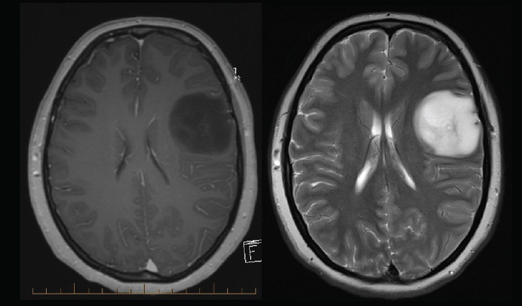
Figure 1. Magnetic resonance imaging showing a typical glioblastoma with central necrosis and peripheral enhancement on contrast-enhanced T1-weighted imaging (left), and surrounding oedema and midline shift on T2-weighted imaging (right)


?Figure 2. Typical low-grade glioma with minimal mass effect

Figure 2. Typical low-grade glioma with minimal mass effect, absence of enhancement on contrast-enhanced T1-weighted magnetic resonance imaging (left), with little surrounding oedema on T2-weighted magnetic resonance imaging (right). The imaging is consistent with numerous diagnoses including astrocytoma and oligodendroglioma, but this case is a World Health Organization grade II diffuse astrocytoma.


Initial management

On diagnosis of a probable brain tumour, the first challenge is breaking the bad news to the patient, keeping in mind that the diagnosis is not certain until histology is obtained. Symptoms of mass effect may be improved using dexamethasone, especially if there is associated cerebral oedema. A typical starting dose is 4 mg stat then daily in the morning. Anticonvulsants are indicated for seizures but not recommended for prophylaxis.1

Urgent neurosurgical review is required for patients with symptoms or signs of raised intracranial pressure, midline shift, hydrocephalus or neurological deficit, and these patients should be referred to an emergency department. Those with a long duration of symptoms and characteristic low-grade or small tumours on MRI may be reviewed as outpatients but should be discussed with a neurosurgeon or neurosurgical registrar.

Surgery

Surgery plays an important part in determining the pathological diagnosis and relieving symptoms. It may improve the efficacy of adjuvant therapy. Both debulking and complete resection of enhancing tumour are associated with extended survival when compared with biopsy alone, but it is not certain whether this association is causative or related to associated tumour and patient factors.5 The benefit of debulking has been confirmed in patients aged >65 years with GBM, and many elderly patients will be considered appropriate for craniotomy.6 Some elderly patients may prefer not to undergo cancer therapy; in this case, supportive palliative care should always be offered as an alternative, particularly for those with poor performance status or multiple comorbidities who benefit least from treatment.

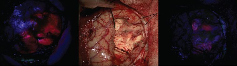
Surgical safety and efficacy are continually improving with modern technology. Fluorescence guidance with 5-aminolevulinic acid (Figure 3) has been shown to improve the extent of resection of high-grade glioma.7 For patients with a tumour near eloquent areas, awake craniotomy with intraoperative neurophysiological monitoring can be used to improve safety.5 Early postoperative MRI is essential to assess residual enhancing tumour and plan radiotherapy. Patients can expect to stay in hospital for approximately five days after an uncomplicated craniotomy, and most need at least six weeks off work.


그렇지 않으면 등급이 낮은 것으로 생각됩니다 .4
그림 1. 대비가 강화 된 T1 가중 이미징 (왼쪽)과 T2 가중 이미징의 주변 부종 및 중간 선 이동에서 중앙 괴사 및 말초 기능 향상이있는 전형적인 교 모세포종을 보여주는 자기 공명 영상 (오른쪽)

-------------------------------------------------- ------------------------------
그림 2. 질량 효과가 가장 적은 전형적인 저급 신경 교종
그림 2. 질량 효과가 최소화되고 대비가 강화 된 T1 가중 자기 공명 영상 (왼쪽)이 향상되지 않은 전형적인 저급 신경 교종 (왼쪽), T2 가중 자기 공명 영상의 주변 부종이 거의 없음 (오른쪽). 영상화는 성상 세포종 및 oligodendroglioma를 포함한 수많은 진단과 일치하지만,이 경우 세계 보건기구 등급 II 확산 성 성상 세포종입니다.

-------------------------------------------------- ------------------------------

초기 관리

가능한 뇌종양을 진단 할 때 첫 번째 과제는 환자에게 나쁜 소식을 전하는 것인데, 조직학을 얻을 때까지 진단이 확실하지 않다는 점을 명심하십시오. 특히 뇌 부종과 관련된 경우 덱사메타손을 사용하여 질량 효과의 증상을 개선 할 수 있습니다. 전형적인 시작 용량은 4 mg 통계량이며 매일 아침입니다. 항 경련제는 발작에 사용되지만 예방에는 권장되지 않습니다 .1

두개 내압, 중추 이동, 뇌수종 또는 신경 학적 결손의 증상 또는 징후가있는 환자의 경우 긴급한 신경 외과 적 검토가 필요하며 이러한 환자는 응급실에 문의해야합니다. 증상이 오래 지속되고 MRI에서 저급 또는 소 종양이 특징 인 환자는 외래 환자로 검토 될 수 있지만 신경 외과 의사 나 신경 외과 레지스트라와상의해야합니다.

수술

병리 진단 및 증상 완화에있어 수술은 중요한 역할을합니다.
Figure 3. Intraoperative imaging of fluorescence-guided surgery using 5-aminolevulinic acid

Figure 3. Intraoperative imaging of fluorescence-guided surgery using 5-aminolevulinic acid. On opening the skull, the tumour can be seen fluorescing pink (left). After tumour resection, under conventional white light microscopy the surgical cavity has no apparent tumour (centre), but further tumour is visible fluorescing under blue light (right) and can be resected.


Diagnosis and molecular advances

Classification of tumours of the central nervous system now requires molecular analysis for characteristic mutations in addition to phenotypic assessment.8 The most important distinction is between gliomas with and without mutations in one of two genes for isocitrate dehydrogenase (IDH). Phenotypically, high-grade gliomas without IDH mutation (IDH wild type) are the most aggressive and correspond to classic GBM. Tumours with the same histological features (ie pleomorphism, necrosis and endothelial proliferation) associated with IDH mutation show a better prognosis and overlap substantially with what was previously designated ‘secondary GBM’.8 Common IDH mutations can be shown on immunohistochemical staining; however, for younger patients or those with other (molecular) features suggestive of IDH mutation, genetic sequencing should be performed if the immunohistochemistry is negative.

The second major division is that in tumours with IDH mutation, co-deletion of chromosome arms 1p and 19q designates oligodendroglioma. IDH mutated/1p19q co-deleted (ie oligodendroglial) tumours have the best prognosis and the best response to treatment.9

Another important molecular marker is methylation of the promotor for the O-6-methylguanine-DNA methyltransferase (MGMT) gene, which is predictive for response to alkylating chemotherapy.9

The recent explosion in molecular data about primary brain tumours has identified numerous other mutations, such as the BRAF V600E mutation in epithelioid GBM and the H3-K27M mutation in diffuse midline glioma, which may be actionable targets for future treatments.8

Concurrent chemoradiation and adjuvant chemotherapy for glioblastoma

The current recommended treatment for GBM is based on the protocol of Stupp et al.1,9 This involves daily temozolomide, an oral alkylating agent, during radiotherapy (60 Gy in 30 fractions over six weeks), followed by six months of temozolomide for five days in 28 days. For older patients, hypofractionated radiotherapy (40 Gy in 15 fractions) has similar efficacy and may be given with or without temozolomide10?12 to reduce treatment time and burden. The target volume includes a 1?2 cm margin around the tumour treated by conformal radiotherapy to avoid vulnerable structures.9,13 Alopecia in the irradiated area, local skin reaction and fatigue that persists beyond radiotherapy are common side effects. Raised intracranial pressure causing headache, nausea and vomiting is possible and can be improved using dexamethasone.

For patients with poor performance status who are aged >70 years and are likely not to tolerate concurrent chemoradiation, temozolomide alone provides similar benefit to radiation alone and may be an appropriate alternative, especially for patients with MGMT promotor methylation.11,12

Temozolomide chemotherapy is usually very well tolerated. The most common adverse reactions are mild-to-moderate nausea and vomiting, transient myelosuppression with thrombocytopenia or neutropenia (~20%), and fatigue that may develop over months.14 Prophylaxis against Pneumocystis jirovecii (previously P. carinii) pneumonia is recommended for patients who continue to take ≥4 mg of dexamethasone during temozolomide treatment.1,14

그림 3. 5-aminolevulinic acid를 사용한 형광 유도 수술의 수술 내 영상 그림 3. 5-aminolevulinic acid를 이용한 형광 유도 수술의 수술 간 영상 두개골을 열면 종양이 분홍색으로 형광이 보입니다 (왼쪽). 종양 절제 후, 통상적 인 백색광 현미경 하에서 수술 공동은 명백한 종양 (중심)을 갖지 않지만, 추가의 종양은 청색광 하에서 형광이 보이고 (오른쪽) 절제 될 수있다.

-------------------------------------------------- ------------------------------

진단 및 분자 발전

중추 신경계의 종양 분류는 표현형 평가에 추가하여 특징적인 돌연변이에 대한 분자 분석이 필요하다. 표현형으로, IDH 돌연변이가없는 고급 신경 교종 (IDH 야생형)이 가장 공격적이고 고전적인 GBM에 해당합니다. IDH 돌연변이와 관련된 조직 학적 특징 (즉, 다형성, 괴사 및 내피 증식)을 갖는 종양은 이전에 '이차 GBM'으로 지정된 것과 더 나은 예후 및 중첩을 나타낸다. 그러나, 젊은 환자 또는 IDH 돌연변이를 암시하는 다른 (분자) 특징을 갖는 환자의 경우, 면역 조직 화학이 음성 인 경우 유전자 시퀀싱을 수행해야한다.

두 번째 주요 부문은 IDH 돌연변이가있는 종양에서 염색체 암 1p 및 19q의 공동 결실이 oligodendroglioma를 지정한다는 것입니다. IDH 돌연변이 / 1p19q 공동-삭제 된 (즉, oligodendroglial) 종양은 치료에 대한 최상의 예후와 최고의 반응을 보인다 .9

또 다른 중요한 분자 마커는 알킬화 화학 요법에 대한 반응을 예측하는 O-6- 메틸 구아닌 -DNA 메틸 트랜스퍼 라제 (MGMT) 유전자에 대한 촉진제의 메틸화입니다 .9

1 차 뇌 종양에 대한 분자 데이터의 최근 폭발은 상피 GBM에서의 BRAF V600E 돌연변이 및 확산 된 중간 선 신경 교종에서의 H3-K27M 돌연변이와 같은 수많은 다른 돌연변이를 확인하였으며, 이는 향후 치료를위한 실행 가능한 표적 일 수있다.

교 모세포종에 대한 동시 화학 방사선과 보조 화학 요법

GBM에 대해 현재 권장되는 치료법은 Stupp et al.1,9의 프로토콜을 기반으로합니다 .1.9 여기에는 방사선 치료 (6 주 동안 30 개 분획에서 60Gy) 후 5 개월 동안 6 개월의 테모 졸로 마이드가 경구 알킬화제 인 일일 테모 졸로 마이드가 포함됩니다. 28 일 만에 고령 환자의 경우, 저 분획 방사선 요법 (15 개 분획에서 40Gy)은 유사한 효능을 가지며, 치료 시간 및 부담을 줄이기 위해 테모 졸로 마이드 10? 12의 유무에 관계없이 제공 될 수 있습니다. 대상 체적은 취약한 구조를 피하기 위해 등각 방사선 요법으로 치료 한 종양 주위에 1-2cm의 마진을 포함합니다 .9,13 방사선 조사 영역에서 탈모증, 국소 피부 반응 및 방사선 요법 이상으로 지속되는 피로는 일반적인 부작용입니다. 두통, 메스꺼움 및 구토를 유발하는 두개 내압 상승이 가능하며 덱사메타손을 사용하여 개선 될 수 있습니다.

70 세 이상이고 동시 화학 방사선 요법을 견딜 수없는 성능 상태가 좋지 않은 환자의 경우 테모 졸로 마이드 단독만으로도 방사선에만 유사한 이점을 제공하며 특히 MGMT 프로모터 메틸화 환자에게 적절한 대안이 될 수 있습니다.

Temozolomide 화학 요법은 일반적으로 매우 잘 견딜 수 있습니다. 가장 흔한 부작용은 경증에서 중등도의 구역 및 구토, 혈소판 감소증 또는 호중구 감소증을 동반 한 일시적 골수 억제 (~ 20 %) 및 수개월에 걸쳐 발생할 수있는 피로입니다 .14 테모 졸로 마이드 치료 동안 덱사메타손 ≥4 mg을 계속 복용하는 환자

Treatment for astrocytomas and oligodendrogliomas

The ideal treatment for lower-grade astrocytomas and oligodendrogliomas (IDH mutated, World Health Organization [WHO] grade II and III) is hotly debated. Patients with a growing, unresectable tumour or uncontrolled seizures should have upfront treatment usually starting with six weeks of radiotherapy (54 Gy).9,15 For other patients, although a significant survival benefit is conferred by radiotherapy and chemotherapy, it is not known whether treatment is better early, as soon as the diagnosis is made, or delayed as long as possible.15?17 The best chemotherapeutic regimen is also unclear. Procarbazine/lomustine/vincristine (PCV) is proven to be effective, particularly for oligodendroglial tumours, but has serious potential side effects including peripheral neuropathy. More recently, temozolomide has been used, supported by retrospective data and the Concurrent and Adjuvant Temozolomide Chemotherapy in Non-1p/19q Deleted Anaplastic Glioma (CATNON) trial for anaplastic (WHO III) astrocytomas.15,17,18 Patients with these tumours should be managed by subspecialist multidisciplinary neuro-oncology teams to make individualised, patient-centred treatment decisions. Ultimately, the choice of timing and modality of treatment will be determined by patient preferences and local practice.

Quality of life and general practice management within the community

The incurable nature of GBM means quality of life during treatment is of the utmost importance. In addition to focal neurological deficits and fatigue ? which may result from the tumour, surgery and radiotherapy ? many patients have insomnia, cognitive problems and psychological issues relating to the diagnosis. Rehabilitation has been shown to be as effective in restoring function for patients with brain tumour as it is in patients following stroke.1,19 Those with symptoms or functional disability from neurological or cognitive impairment may benefit from referral to appropriate allied health services, including occupational therapy, speech pathology and neuropsychology, whether or not the patient has had surgery.

Ongoing steroids may be required to manage symptoms and radiotherapy-related oedema but should be weaned as soon as possible in consultation with an oncologist. Patients should be monitored for side effects, including diabetes, infections and skin lesions, and weaning should be carried out gradually to avoid symptomatic adrenal insufficiency.14 First-line anticonvulsants for treatment of seizures include sodium valproate for generalised seizures and carbamazepine for focal events. In patients who have not had seizures, anticonvulsants can be stopped after surgery.1,9

Depressive symptoms are common and may be addressed by psychosocial support, cognitive behavioural therapy or mindfulness training. A referral to a psychologist may be helpful.9,20 Continuing moderate regular exercise is beneficial, but support and direction from an exercise physiologist or physiotherapist may be necessary.21

Driving is proscribed for six months after supratentorial surgery,22 but seizures and cognitive or neurological deficits prevent many patients with glioma regaining their licence. If in doubt, an occupational therapy driving assessment is essential.

Psychosocial issues are particularly important for patients with low-grade glioma who may live for years or decades after diagnosis and are often younger, with dependent family and financial obligations. GPs can provide an important educational and supportive role as patients grapple with their responsibilities and self-identity in the face of the existential threat and possible impairment and personality change caused by a tumour. The family is also likely to benefit from counselling and support. Online resources (Table 1) and local Cancer Council support groups are often useful.

성상 세포종 및 oligodendrogliomas의 치료

저급 성상 세포종 및 소 생식 종증 (IDH 돌연변이, 세계 보건기구 (WHO) 등급 II 및 III)에 대한 이상적인 치료법에 대한 논쟁이 뜨겁습니다. 커지고, 절제 할 수없는 종양이나 통제되지 않은 발작이있는 환자는 일반적으로 6 주간의 방사선 요법 (54 Gy)으로 시작하여 선행 치료를 받아야합니다. 진단이 이루어 지거나 가능한 한 빨리 치료하는 것이 더 좋습니다. 15? 17 최고의 화학 요법은 확실하지 않습니다. 프로 카바 진 / 로무 스틴 / 빈 크리스틴 (PCV)은 특히 oligodendroglial 종양에 효과적인 것으로 입증되었지만 말초 신경 병증을 포함한 심각한 잠재적 부작용이 있습니다. 가장 최근에, 테모 졸로 마이 드는 회고 적 데이터 및 역 형성 (WHO III) 성상 세포종에 대한 비 -1p / 19q 결실 된 해부학 적 신경 교종 (CATNON) 시험에서의 동시 및 보조적 테모 졸로 마이드 화학 요법에 의해지지되어왔다 .15,17,18 하위 전문의 다 분야 신경 종양학 팀에 의해 관리되어 개별화 된 환자 중심 치료 결정을 내립니다. 궁극적으로, 치료시기 및 치료 방식의 선택은 환자의 선호 및 현지 진료에 의해 결정될 것이다.

지역 사회 내 삶의 질 및 일반적인 관행 관리

GBM의 치료가 불가능한 특성은 치료 중 삶의 질이 가장 중요하다는 것을 의미합니다. 종양, 수술 및 방사선 요법으로 인해 발생할 수있는 국소 신경 학적 결손 및 피로 외에도 많은 환자들이 진단과 관련하여 불면증,인지 문제 및 심리적 문제가 있습니다. 재활은 뇌졸중 환자와 마찬가지로 뇌종양 환자의 기능을 회복시키는 데 효과적인 것으로 나타났습니다. 환자가 수술을 받았는지 여부에 관계없이 요법, 언어 병리학 및 신경 심리학.

진행중인 스테로이드는 증상 및 방사선 치료 관련 부종을 관리하는 데 필요할 수 있지만 종양 전문의와 상담하여 가능한 한 빨리 쇠약해야합니다. 환자는 당뇨병, 감염 및 피부 병변을 포함한 부작용에 대해 모니터링해야하고 증상 부신 기능 부전을 피하기 위해 점차적으로 이유를 수행해야합니다 .14 발작 치료를위한 1 차 항 경련제로는 발작 치료를위한 나트륨 발 프로 에이트와 국소 증상에 대한 카르 바 마제 핀이 있습니다. 발작이없는 환자의 경우 수술 후 항 경련제가 멈출 수 있습니다 .1,9

우울 증상은 일반적이며 심리 사회적 지원,인지 행동 요법 또는 마음 챙김 훈련으로 해결할 수 있습니다. 심리학자에게 의뢰하는 것이 도움이 될 수 있습니다 .9,20 계속적인 규칙적인 운동은 유익하지만 운동 물리학 자나 물리 치료사의 도움과 지시가 필요할 수 있습니다 .21

상복 수술 후 6 개월 동안 운전이 허용되지만, 발작과인지 적 또는 신경 학적 결함은 신경 교종 환자가 면허증을 되 찾는 것을 막습니다. 의심스러운 경우, 직업 요법 운전 평가가 필수적입니다.

심리 사회적 문제는 진단 후 수십 년 또는 수십 년 동안 살 수 있고 종종 가족과 재정적 의무가있는 어린 나이의 저급 신경 교종 환자에게 중요합니다. GP는 환자가 존재하는 위협과 종양으로 인한 가능한 손상 및 성격 변화에 직면하여 자신의 책임과 자기 정체성을 파악함에 따라 중요한 교육 및 지원 역할을 제공 할 수 있습니다. 가족은 또한 상담과 지원으로 혜택을 볼 수 있습니다. 온라인 자료 (표 1)와 현지 암 협의회 지원 그룹이 종종 유용합니다.

Management of recurrent tumour

Even with maximal therapy, GBM tumours almost inevitably return. Despite this, there is no standard of care for management of progression. Re-operation is useful to distinguish recurrence from treatment effect (pseudoprogression) and is associated with longer survival, but this may be due to sel ection bias. In some cases, re-irradiation is feasible. Second-line chemotherapy options include metronomic temozolomide (continuous daily instead of five days in 28) and lomustine.

Bevacizumab has recently been approved on the Pharmaceutical Benefits Scheme for use in recurrent or progressive GBM. This monoclonal antibody targets vascular endothelial growth factor, a gene commonly overexpressed in GBM. Trial evidence to date suggests that bevacizumab restores the blood?brain barrier, reducing enhancement on MRI scans and reducing oedema, thereby improving symptoms. However, bevacizumab does not have cytotoxic effects that extend survival.9

Tumour treating fields are a novel treatment modality using alternating electrical fields applied to the head through electrodes worn for 18 hours per day.9 The mechanism for the supposed effect remains unclear; there is no blinded randomised controlled trial and it requires purchase of expensive disposables, causing scepticism among many neuro-oncologists.23 It is unlikely to be introduced to Australia in the foreseeable future.

Current research frontiers

Unlike trastuzumab for breast cancer, sunitinib for renal cancer and dabrafenib for melanoma, no targeted agents have yet been identified that improve survival in GBM. Research continues, with numerous Australian trials currently underway, including trials investigating agents active against EphA3 receptors (KB004), tubulin (ACT001) and transcription factors PAX-1 and NF-kB. Many guidelines recommend enrolment in a clinical trial as optimal care for glioma patients.1,9,24

Although the brain is relatively protected from systemic immune responses, there is laboratory and clinical evidence that immunotherapy may be harnessed against brain tumours by counteracting the tumour-induced immunosuppression (immunomodulation), sensitising immune cells to tumour antigens (tumour vaccines) or by in vitro expansion and reinfusion of anti-tumour immune cells (adoptive immunotherapy). The Australian NUTMEG trial is currently testing immunomodulation with the PD-1 checkpoint inhibitor nivolumimab in patients aged >65 years with unmethylated GBM, and more than a dozen similar trials are underway overseas. Tumour vaccines and adoptive immunotherapy have to date been unsuccessful, but this remains an active field of research.25

재발 성 종양 관리

최대 요법으로도 GBM 종양은 거의 필연적으로 돌아옵니다. 그럼에도 불구하고, 진행 관리를위한 표준은 없습니다. 재수술은 재발을 치료 효과 (의사 진행)와 구별하는 데 유용하며 생존 기간이 길어 지지만, 이는 선입 편향 때문일 수 있습니다. 어떤 경우에는 재조사가 가능합니다. 2 차 화학 요법 옵션에는 메트로놈 테모 졸로 마이드 (28 일 5 일 대신 연속) 및 로무 스틴이 포함됩니다.

베바 시주 맙은 최근 재발 성 또는 점진적 GBM에 사용하기위한 제약 혜택 제도에 대해 승인되었습니다. 이 단일 클론 항체는 GBM에서 일반적으로 과발현되는 유전자 인 혈관 내피 성장 인자를 표적으로한다. 현재까지의 시험 증거는 베바 시주 맙이 혈액-뇌 장벽을 회복시켜 MRI 스캔의 향상을 감소시키고 부종을 감소시켜 증상을 개선 시킨다는 것을 시사한다. 그러나 베바 시주 맙에는 생존을 연장시키는 세포 독성 효과가 없습니다 .9

종양 치료 분야는 하루에 18 시간 동안 착용 된 전극을 통해 머리에 가해진 교류 전기장을 사용하는 새로운 치료 방식입니다 .9 가정 된 효과의 메커니즘은 불분명합니다. 맹검 무작위 배정 시험은 없으며, 많은 신경 종양 학자들 사이에 회의론을 유발하는 고가의 일회용 제품을 구입해야합니다 .23 가까운 미래에 호주에 소개 될 것 같지 않습니다.

현재 연구 경계

유방암에 대한 트라 스투 주맙, 신장 암에 대한 수 니티 닙 및 흑색 종에 대한 다 브라 페닙과는 달리 GBM의 생존을 개선시키는 표적 제는 아직 밝혀지지 않았다. EphA3 수용체 (KB004), 튜 불린 (ACT001) 및 전사 인자 PAX-1 및 NF-kB에 대해 활성 물질을 조사하는 시험을 포함하여 수많은 호주 시험이 현재 진행되고 있습니다. 많은 가이드 라인은 신경 교종 환자를위한 최적의 치료법으로 임상 시험에 등록 할 것을 권장합니다 .1,9,24

뇌는 전신 면역 반응으로부터 비교적 보호되지만, 종양 유발 면역 억제 (면역 조절), 면역 세포를 종양 항원 (종양 백신)에 민감하게하거나 시험관 내에서 면역 치료가 ??뇌종양에 대해 이용 될 수 있다는 실험실 및 임상 증거가 있습니다. 항-종양 면역 세포의 확장 및 재 주입 (입양 면역 요법). 호주 NUTMEG 시험은 현재 메틸화되지 않은 GBM을 가진 65 세 초과 환자에서 PD-1 체크 포인트 억제제 니볼 루미 맙으로 면역 조절을 시험하고 있으며, 12 개 이상의 유사한 시험이 해외에서 진행되고있다. 종양 백신과 입양 면역 요법은 현재까지 성공하지 못했지만 이것은 활발한 연구 분야로 남아 있습니다 .25

The use of modified viruses to activate the immune system against the tumour cells is also under investigation. In a Phase II dose-finding study of a recombinant non-pathogenic polio-rhinovirus chimera (PVSRIPO) infused into the brain by convection-enhanced delivery, 20% of patients remain alive and well three years after treatment for recurrent GBM.26 Similar but less spectacular results have been observed after trials of other viral treatments.26 This offers hope that oncolytic viral therapy may provide lasting benefit for at least some patients, although optimism should be tempered as many treatments that appear promising in Phase II trials have been ineffective in large randomised controlled Phase III trials.27

Prognosis

Patients vary in their desire to discuss prognosis, and statistical averages do not necessarily apply to a particular patient. The median survival of patients aged >65 years with GBM is under three months after biopsy alone,6 whereas for younger patients with compete resection of enhancement, median survival is nearly two years.5 This means half the patients may live longer than two years. Population data suggest the five-year survival for patients with GBM is approaching 10%, a significant improvement on earlier decades.28

Lower-grade gliomas have an even more variable prognosis because of the unpredictable progression to high-grade tumours. Median survival after radiation and PCV chemotherapy for diffuse WHO grade II astrocytomas and oligodendrogliomas is >13 years.16

Palliative care

Referral to specialist palliative care services is valuable for patients with high-grade glioma as soon as the patient has needs that these services may address. Early referral enables relationships to be established and plans made before the terminal phase of the illness, and is associated with fewer unplanned hospital admissions and better symptom control.1,9 Terminal palliative care undertaken by GPs within the patient’s local community enables the best possible quality of life for patients with brain tumours during their final months.

종양 세포에 대한 면역계를 활성화시키기 위해 변형 된 바이러스의 사용이 또한 조사 중에있다. 대류-강화 전달에 의해 뇌에 주입 된 재조합 비병원성 폴리오-리노 바이러스 키메라 (PVSRIPO)의 II 상 용량-조사 연구에서, 환자의 20 %가 재발하는 GBM 치료 후 3 년 후에 생존하고 잘 유지된다 .26 유사하지만 다른 바이러스 치료를 시행 한 후에도 눈에 띄지 않는 결과가 관찰되었습니다 .26 이것은 종양 용해성 바이러스 치료가 적어도 일부 환자에게 지속적인 혜택을 제공 할 수 있기를 희망하지만, 제 2 상 임상 시험에서 유망한 것으로 보이는 많은 치료법이 효과가 없어 낙관주의가 완화되어야하지만 대규모 무작위 통제 된 3 상 임상 시험 .27

예지

환자는 예후에 대해 논의하고자하는 바가 다양하며, 통계적 평균이 특정 환자에게 반드시 적용되는 것은 아닙니다. GBM을 가진 65 세 초과 환자의 평균 생존율은 생검 단독으로 3 개월 미만이지만 6, 경쟁 강화 절제술을받은 젊은 환자의 경우 평균 생존율은 거의 2 년입니다 .5 이는 환자의 절반이 2 년 이상 생존 할 수 있음을 의미합니다. 인구 데이터에 따르면 GBM 환자의 5 년 생존율이 10 %에 가까워지고 있습니다.

낮은 등급의 신경 교종은 예측할 수없는 고급 종양으로의 진행으로 인해 더욱 다양한 예후를 보입니다. 확산 WHO 등급 II 성상 세포종 및 소 생식 종증에 대한 방사선 및 PCV 화학 요법 후 평균 생존율은> 13 년이다 .16

완화 치료

전문적인 완화 치료 서비스에 대한 의뢰는 환자가 이러한 서비스로 해결할 수있는 요구가있는 즉시 고급 신경 교종 환자에게 유용합니다. 조기 진료 의뢰는 질병의 말기 단계 이전에 관계를 수립하고 계획을 세우고 계획되지 않은 병원 입원 횟수를 줄이고 증상을보다 효과적으로 통제 할 수 있도록합니다 .1,9 환자의 지역 사회 내 GP가 수행하는 말기 완화 치료는 최상의 품질을 가능하게합니다. 마지막 몇 달 동안 뇌종양 환자의 삶의 질.

Key points

  • Management of patients with primary brain tumours is complex, so patients should be referred to a tertiary centre with subspecialist neuro-oncologists and a brain tumour multidisciplinary team.
  • Recent advances in the molecular understanding of brain tumours have improved prognostication but not yet changed treatment significantly.
  • Primary care physicians can ameliorate the burden of illness by managing the significant symptoms, psychosocial issues and supportive care needs encountered by patients with primary brain tumours.
Competing interests: None.
Provenance and peer review: Commissioned, externally peer reviewed.
Funding: None.
References
  1. Australian Cancer Network Adult Brain Tumour Guidelines Working Party. Clinical practice guidelines for the management of adult gliomas: Astrocytomas and oligodendrogliomas. Sydney, NSW: Cancer Council Australia, Australian Cancer Network and Clinical Oncology Society of Australia Inc, 2009. Search PubMed
  2. Ozawa M, Brennan PM, Zienius K, et al. The usefulness of symptoms alone or combined for general practitioners in considering the diagnosis of a brain tumour: A case-control study using the clinical practice research database (CPRD) (2000-2014). BMJ Open 2019;9(8):e029686. doi: 10.1136/bmjopen-2019-029686. Search PubMed
  3. Shaw TB, Jeffree RL, Thomas P, et al. Diagnostic performance of 18F-fluorodeoxyglucose positron emission tomography in the evaluation of glioma. J Med Imaging Radiat Oncol 2019;63(5):650?56. doi: 10.1111/1754-9485.12929. Search PubMed
  4. Chukwueke UN, Wen PY. Use of the response assessment in neuro-oncology (RANO) criteria in clinical trials and clinical practice. CNS Oncol 2019;8(1):CNS28. doi: 10.2217/cns-2018-0007. Search PubMed
  5. D’Amico RS, Englander ZK, Canoll P, Bruce JN. Extent of resection in glioma ? A review of the cutting edge. World Neurosurg 2017;103:538?49. doi: 10.1016/j.wneu.2017.04.041. Search PubMed
  6. Vuorinen V, Hinkka S, F?rkkil? M, J??skel?inen J. Debulking or biopsy of malignant glioma in elderly people ? A randomised study. Acta Neurochir (Wien) 2003;145(1):5?10. doi: 10.1007/s00701-002-1030-6 Search PubMed
  7. Colditz MJ, Jeffree RL. Aminolevulinic acid (ALA)-protoporphyrin IX fluorescence guided tumour resection. Part 1: Clinical, radiological and pathological studies. J Clin Neurosci 2012;19(11):1471?74. doi: 10.1016/j.jocn.2012.03.009. Search PubMed
  8. Wesseling P, Capper D. WHO 2016 Classification of gliomas. Neuropathol Appl Neurobiol 2018;44(2):139?50. doi: 10.1111/nan.12432. Search PubMed
  9. National Comprehensive Cancer Network. Central nervous system cancers. Plymouth Meeting: NCCN, 2019. Search PubMed
  10. Roa W, Brasher PM, Bauman G, et al. Abbreviated course of radiation therapy in older patients with glioblastoma multiforme: A prospective randomized clinical trial. J Clin Oncol 2004;22(9):1583?88. doi: 10.1200/JCO.2004.06.082. Search PubMed
  11. Malmstr?m A, Grønberg BH, Marosi C, et al. Temozolomide versus standard 6-week radiotherapy versus hypofractionated radiotherapy in patients older than 60 years with glioblastoma: The Nordic randomised, phase 3 trial. Lancet Oncol 2012;13(9):916?26. doi: 10.1016/S1470-2045(12)70265-6. Search PubMed
  12. 키 포인트
    원발성 뇌종양 환자의 관리는 복잡하므로, 환자는 전문의 신경 종양 전문의와 뇌종양 다방면 팀을 갖춘 3 차 센터에 문의해야합니다.
    뇌종양의 분자 이해에 대한 최근의 진보는 예후는 개선되었지만 아직 치료를 크게 변화 시키지는 않았다.
    1 차 진료 의사는 1 차 뇌종양 환자가 겪는 심각한 증상, 심리 사회적 문제 및 지원 적 치료 요구를 관리함으로써 질병의 부담을 완화 할 수 있습니다.

    경쟁 관계 : 없음

    Provenance and Peer Review : 위탁, 외부 동료 검토.

    자금 : 없음.


    참고 문헌
    호주 암 네트워크 성인 뇌종양 지침 작업반. 성인 신경 교종 관리를위한 임상 실습 지침 : Astrocytomas 및 oligodendrogliomas. 시드니, 뉴 사우스 웨일즈 : 호주 암 협의회, 호주 암 네트워크 및 호주 임상 암 학회, 2009. PubMed 검색
    Ozawa M, Brennan PM, Zienius K 등 뇌종양 진단을 고려할 때 일반 개업의에게 단독으로 또는 병용 된 증상의 유용성 : 임상 실습 연구 데이터베이스 (CPRD)를 이용한 사례 관리 연구 (2000-2014). BMJ 오픈 2019; 9 (8) : e029686. 도 : 10.1136 / bmjopen-2019-029686. PubMed 검색
    Shaw TB, Jeffree RL, Thomas P 등 신경 교종의 평가에서 18F- 플루오로 데 옥시 글루코스 양전자 방출 단층 촬영의 진단 성능. J Med Imaging Radiat Oncol 2019; 63 (5) : 650? 56. 도 : 10.1111 / 1754-9485.12929. PubMed 검색
    Chukwueke UN, Wen PY. 임상 시험 및 임상 실습에서 신경 종양학 (RANO) 기준에서 반응 평가의 사용. CNS Oncol 2019; 8 (1) : CNS28. 도 : 10.2217 / cns-2018-0007. PubMed 검색
    D' Amico RS, Englander ZK, Canoll P, Bruce JN. 신경 교종의 절제 범위 ― 최첨단 검토. World Neurosurg 2017; 103 : 538? 49. 도 : 10.1016 / j.wneu.2017.04.041. PubMed 검색
    Vuorinen V, Hinkka S, F? rkkil? M, J ?? skel? inen J. 노인에서 악성 신경 교종의 디 벌킹 또는 생검 ™ 무작위 연구. Acta Neurochir (Wien) 2003; 145 (1) : 5? 10. doi : 10.1007 / s00701-002-1030-6 검색 PubMed
    Colditz MJ, Jeffree RL. 아미노 레 불린 산 (ALA)-프로토 포르피린 IX 형광 유도 종양 절제. 1 부 : 임상, 방사선 및 병리학 연구. J Clin Neurosci 2012; 19 (11) : 1471? 74. 도 : 10.1016 / j.jocn.2012.03.009. PubMed 검색
    Wesseling P, Capper D. WHO 2016 신경 교종 분류. Neuropathol Appl Neurobiol 2018; 44 (2) : 139? 50. 도 : 10.1111 / nan.12432 PubMed 검색
    전국 종합 암 네트워크. 중추 신경계 암. 플리머스 회의 : 2019 년 NCCN. PubMed 검색
    Roa W, Brasher PM, Bauman G 등 다형성 교 모세포종 환자에서 방사선 요법의 약식 과정 : 전향 적 무작위 임상 시험. J Clin Oncol 2004; 22 (9) : 1583? 88. 도 : 10.1200 / JCO.2004.06.082. PubMed 검색
    Malmstr? m A, Grønberg BH, Marosi C, 등. 교 모세포종이있는 60 세 이상의 환자에서 테모 졸로 마이드 대 표준 6 주 방사선 요법 대 저 분획 방사선 요법 : 노르딕 무작위, 3 상 시험. Lancet Oncol 2012; 13 (9) : 916? 26. 도 : 10.1016 / S1470-2045 (12) 70265-6. PubMed 검색
    1. Mann J, Ramakrishna R, Magge R, Wernicke AG. Advances in radiotherapy for glioblastoma. Front Neurol 2018;8:748. doi: 10.3389/fneur.2017.00748. Search PubMed
    2. Louis DN, Perry A, Reifenberger G, et al. The 2016 World Health Organization classification of tumors of the central nervous system: A summary. Acta Neuropathol 2016;131(6):803?20. doi: 10.1007/s00401-016-1545-1. Search PubMed
    3. MIMS. MIMS online. NSW, Australia: North Sydney, NSW: MIMS Australia Pty Ltd, 2019. Available at www.mimsonline.com.au [Accessed 13 December 2019]. Search PubMed
    4. National Institute for Health and Care Excellence. Brain tumours (primary) and brain metastases in adults. NICE guideline [NG99]. London: NICE, 2018 Search PubMed
    5. Ruff MW, Buckner JC, Johnson DR, van den Bent MJ, Geurts M. Neuro-oncology clinical debate: PCV or temozolomide in combination with radiation for newly diagnosed high-grade oligodendroglioma. Neurooncol Pract 2019;6(1):17?21. doi: 10.1093/nop/npy044. Search PubMed
    6. van den Bent MJ, Afra D, de Witte O, et al. Long-term efficacy of early versus delayed radiotherapy for low-grade astrocytoma and oligodendroglioma in adults: The EORTC 22845 randomised trial. Lancet 2005;366(9490):985?90. doi: 10.1016/S0140-6736(05)67070-5. Search PubMed
    7. van den Bent MJ, Baumert B, Erridge SC, Vogelbaum MA, Nowak AK, Sanson M, et al. Interim results from the CATNON trial (EORTC study 26053-22054) of treatment with concurrent and adjuvant temozolomide for 1p/19q non-co-deleted anaplastic glioma: a phase 3, randomised, open-label intergroup study. Lancet 2017;390(10103):1645?53. doi: 10.1016/S0140-6736(17)31442-3. Search PubMed
    8. Vargo M, Henriksson R, Salander P. Rehabilitation of patients with glioma. Handb Clin Neurol 2016;134:287?304. doi: 10.1016/B978-0-12-802997-8.00017-7. Search PubMed
    9. Siegel C, Armstrong TS. Nursing guide to management of major symptoms in patients with malignant glioma. Semin Oncol Nurs 2018;34(5):513?27. doi: 10.1016/j.soncn.2018.10.014. Search PubMed
    10. Cormie P, Nowak AK, Chambers SK, Galv?o DA, Newton RU. The potential role of exercise in neuro-oncology. Front Oncol 2015;5:85. doi: 10.3389/fonc.2015.00085. Search PubMed
    11. Austroads. Assessing fitness to drive: For commercial and private vehicle drivers. Sydney, NSW: Austroads, 2016. Search PubMed
    12. Wick W. TTFields: Where does all the skepticism come from? Neuro Oncol 2016;18(3):303?05. doi: 10.1093/neuonc/now012. Search PubMed
    13. High Grade Glioma Working Group NCERG. Optimal cancer care pathway for people with high-grade glioma. Sydney, NSW: Cancer Australia, 2016. Search PubMed
    14. Mann J, Ramakrishna R, Magge R, Wernicke AG. Advances in radiotherapy for glioblastoma. Front Neurol 2018;8:748. doi: 10.3389/fneur.2017.00748. Search PubMed
      Louis DN, Perry A, Reifenberger G, et al. The 2016 World Health Organization classification of tumors of the central nervous system: A summary. Acta Neuropathol 2016;131(6):803?20. doi: 10.1007/s00401-016-1545-1. Search PubMed
      MIMS. MIMS online. NSW, Australia: North Sydney, NSW: MIMS Australia Pty Ltd, 2019. Available at www.mimsonline.com.au [Accessed 13 December 2019]. Search PubMed
      National Institute for Health and Care Excellence. Brain tumours (primary) and brain metastases in adults. NICE guideline [NG99]. London: NICE, 2018 Search PubMed
      Ruff MW, Buckner JC, Johnson DR, van den Bent MJ, Geurts M. Neuro-oncology clinical debate: PCV or temozolomide in combination with radiation for newly diagnosed high-grade oligodendroglioma. Neurooncol Pract 2019;6(1):17?21. doi: 10.1093/nop/npy044. Search PubMed
      van den Bent MJ, Afra D, de Witte O, et al. Long-term efficacy of early versus delayed radiotherapy for low-grade astrocytoma and oligodendroglioma in adults: The EORTC 22845 randomised trial. Lancet 2005;366(9490):985?90. doi: 10.1016/S0140-6736(05)67070-5. Search PubMed
      van den Bent MJ, Baumert B, Erridge SC, Vogelbaum MA, Nowak AK, Sanson M, et al. Interim results from the CATNON trial (EORTC study 26053-22054) of treatment with concurrent and adjuvant temozolomide for 1p/19q non-co-deleted anaplastic glioma: a phase 3, randomised, open-label intergroup study. Lancet 2017;390(10103):1645?53. doi: 10.1016/S0140-6736(17)31442-3. Search PubMed
      Vargo M, Henriksson R, Salander P. Rehabilitation of patients with glioma. Handb Clin Neurol 2016;134:287?304. doi: 10.1016/B978-0-12-802997-8.00017-7. Search PubMed
      Siegel C, Armstrong TS. Nursing guide to management of major symptoms in patients with malignant glioma. Semin Oncol Nurs 2018;34(5):513?27. doi: 10.1016/j.soncn.2018.10.014. Search PubMed
      Cormie P, Nowak AK, Chambers SK, Galv?o DA, Newton RU. The potential role of exercise in neuro-oncology. Front Oncol 2015;5:85. doi: 10.3389/fonc.2015.00085. Search PubMed
      Austroads. Assessing fitness to drive: For commercial and private vehicle drivers. Sydney, NSW: Austroads, 2016. Search PubMed
      Wick W. TTFields: Where does all the skepticism come from? Neuro Oncol 2016;18(3):303?05. doi: 10.1093/neuonc/now012. Search PubMed
      High Grade Glioma Working Group NCERG. Optimal cancer care pathway for people with high-grade glioma. Sydney, NSW: Cancer Australia, 2016. Search PubMed
    1. Mann J, Ramakrishna R, Magge R, Wernicke AG. 교 모세포종에 대한 방사선 치료의 발전. 프론트 뉴롤 2018; 8 : 748. 도 : 10.3389 / fneur.2017.00748. PubMed 검색
      Louis DN, Perry A, Reifenberger G 등 중추 신경계 종양의 2016 세계 보건기구 분류 : 요약. Acta Neuropathol 2016; 131 (6) : 803? 20. 도 : 10.1007 / s00401-016-1545-1. PubMed 검색
      MIMS. MIMS 온라인. NSW, 호주 : 노스 시드니, NSW : MIMS Australia Pty Ltd, 2019. www.mimsonline.com.au에서 이용 가능 [2019 년 12 월 13 일 액세스]. PubMed 검색
      국립 보건 및 의료 연구소. 성인의 뇌종양 (1 차) 및 뇌 전이. 좋은 지침 [NG99]. London : NICE, 2018 Search PubMed
      Ruff MW, Buckner JC, Johnson DR, van den Bent MJ, Geurts M. Neuro-oncology 임상 토론 : 새로 진단 된 고급 oligodendroglioma를위한 방사선과 결합 된 PCV 또는 temozolomide. Neurooncol Pract 2019; 6 (1) : 17? 21. doi : 10.1093 / nop / npy044. PubMed 검색
      van den Bent MJ, Afra D, de Witte O 등 성인의 저급 성상 세포종 및 oligodendroglioma에 대 한 조기 대 지연 방사선 치료의 장기 효능 : EORTC 22845 무작위 시험. Lancet 2005; 366 (9490) : 985? 90. 도 : 10.1016 / S0140-6736 (05) 67070-5. PubMed 검색
      van den Bent MJ, Baumert B, Erridge SC, Vogelbaum MA, Nowak AK, Sanson M 등 1p / 19q 비-코-삭제 결실 역 형성 신경 교종에 대한 동시 및 보조 테모 졸로 마이 드로의 치료의 CATNON 시험 (EORTC 연구 26053-22054)의 중간 결과 : 3 상 무작위 배정, 오픈-라벨 그룹 간 연구. Lancet 2017; 390 (10103) : 1645? 53. 도 : 10.1016 / S0140-6736 (17) 31442-3. PubMed 검색
      Vargo M, Henriksson R, 신경 교종 환자의 Salander P. 재활 Handb Clin Neurol 2016; 134 : 287? 304. 도 : 10.1016 / B978-0-12-802997-8.00017-7. PubMed 검색
      Siegel C, 암스트롱 TS. 악성 신경아 교종 환자의 주요 증상 관리를위한 간호 안내서. Semin Oncol Nurs 2018; 34 (5) : 513? 27. 도 : 10.1016 / j.soncn.2018.10.014. PubMed 검색
      Cormie P, Nowak AK, Chambers SK, Galv? DA, Newton RU. 신경 종양학에서 운동의 잠재적 역할. Front Oncol 2015; 5 : 85. 도 : 10.3389 / fonc.2015.00085. PubMed 검색
      호주. 운전 적합성 평가 : 상용 및 개인 차량 운전자에게 적합합니다. Sydney, NSW : Austroads, 2016. PubMed 검색
      Wick W. TTFields : 모든 회의론은 어디에서 왔습니까? Neuro Oncol 2016; 18 (3) : 303? 05. 도 : 10.1093 / neuonc / now012. PubMed 검색
      고급 신경아 교종 실무 그룹 NCERG. 고급 신경 교종 환자를위한 최적의 암 치료 경로. Sydney, NSW : Cancer Australia, 2016. PubMed 검색
    2. Swartz AM, Shen SH, Salgado MA, Congdon KL, Sanchez-Perez L. Promising vaccines for treating glioblastoma. Expert Opin Biol Ther 2018;18(11):1159?70. doi: 10.1080/14712598.2018.1531846. Search PubMed
    3. Chiocca EA, Nassiri F, Wang J, Peruzzi P, Zadeh G. Viral and other therapies for recurrent glioblastoma: Is a 24-month durable response unusual? Neuro Oncol 2019;21(1):14?25. doi: 10.1093/neuonc/noy170. Search PubMed
    4. Weller M, Butowski N, Tran DD, et al. Rindopepimut with temozolomide for patients with newly diagnosed, EGFRvIII-expressing glioblastoma (ACT IV): A randomised, double-blind, international phase 3 trial. Lancet Oncol 2017;18(10):1373?85. doi: 10.1016/S1470-2045(17)30517-X. Search PubMed
    5. Trada Y, Jeffree RL, Inglis P, Fay M, Guan T. Management of glioblastoma patient in Queensland. Sydney: COGNO Annual Scientific Meeting, October 2013. Search PubMed
    6. Swartz AM, Shen SH, Salgado MA, Congdon KL, Sanchez-Perez L. 교 모세포종 치료를위한 유망한 백신. Expert Opin Biol Ther 2018; 18 (11) : 1159? 70. 도 : 10.1080 / 14712598.2018.1531846. PubMed 검색
      재발 성 교 모세포종에 대한 Chiocca EA, Nassiri F, Wang J, Peruzzi P, Zadeh G. Viral 및 기타 요법 : 24 개월 지속되는 반응이 특이합니까? Neuro Oncol 2019; 21 (1) : 14? 25. 도 : 10.1093 / neuonc / noy170. PubMed 검색
      Weller M, Butowski N, Tran DD 등 새로 진단 된 EGFRvIII- 발현 교 모세포종 (ACT IV) 환자를위한 테모 졸로 마이드를 사용한 린 도피 무트 : 무작위 이중 맹검 국제 3 상 시험. Lancet Oncol 2017; 18 (10) : 1373? 85. 도 : 10.1016 / S1470-2045 (17) 30517-X. PubMed 검색
      Trada Y, Jeffree RL, Inglis P, Fay M, Guan T. 퀸즐랜드의 교 모세포종 환자 관리. 시드니 : COGNO 연례 과학 회의, 2013 년 10 월. PubMed 검색


      https://www1.racgp.org.au/ajgp/2020/april/current-management-of-cerebral-gliomas